Cryo-Electron Microscope
Usage Guide
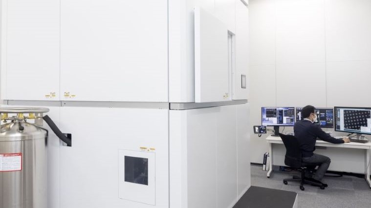
In 2021, Tohoku University Advanced Research Center for Innovations in Next-Generation Medicine (INGEM), with the support of the Japan Agency for Medical Research and Development (AMED) under the Platform Project for Supporting Drug Discovery and Life Science Research, Basis for Supporting Innovative Drug Discovery and Life Science Research (BINDS, https://www.binds.jp/), installed the latest 300 kV field emission cryo-electron microscope (CRYO ARM™ 300 II, manufactured by JEOL Ltd.) equipped with a direct detection camera (K3™, manufactured by GATAN Inc.). Additionally, in 2024, with the continued support from AMED-BINDS, a scanning electron microscope (CRYO-FIB-SEM JIB-4700F, manufactured by JEOL Ltd.) was also installed. At INGEM, these devices are broadly available to researchers both within and outside the university, providing comprehensive support from the preparation of frozen samples to imaging.
What is Cryo-Electron Microscopy?
High-resolution structural information on target proteins is essential for drug discovery based on molecular structures of proteins. Cryo-Electron Microscopy (Cryo-EM) is a technique that rapidly freezes biological samples, trapping them in glassy ice, and directly observes them using a transmission electron microscope. This method allows for the visualization of three-dimensional structures of biological macromolecular complexes at high resolution. Since the process does not require crystallization, it enables the structural analysis of many proteins that are difficult to crystallize for X-ray crystallography. With this device, structural analysis of diverse biological macromolecules at high resolution is possible, providing a molecular basis for cutting-edge life science research and drug discovery.
Furthermore, cryo-electron tomography analysis, which can capture near-living cell states by rapidly freezing cells, has recently attracted attention. Cells to be observed must be sectioned using a focused ion beam (FIB)-scanning electron microscope (SEM) after freezing. At INGEM, the introduction of a new CRYO-FIB-SEM in 2024 has enabled cryo-electron tomography analysis in combination with the CRYO ARM™ 300 II.
About the Equipment
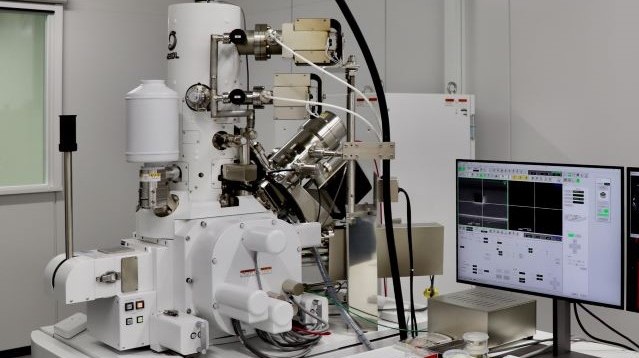
Cryo-Transmission Electron Microscope
CRYO ARM™ 300 II (manufactured by JEOL Ltd.)
Acceleration Voltage: 300 kV, Cold Cathode Field Emission Electron Gun
Direct Detection Camera: K3™ (manufactured by GATAN Inc.)
Cryo-Scanning Electron Microscope
CRYO-FIB-SEM: JIB-4700F (manufactured by JEOL Ltd.)
Cryo Stage: VCT500 (manufactured by Leica Inc.)
Cryo-Fluorescence Microscope System (Cryo Stage: Linkam Scientific Instruments Inc., Fluorescence Microscope: Nikon Solutions Inc.)
Supercomputer System
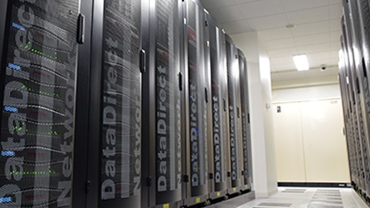
Analysis using the supercomputer installed at the Tohoku Medical Megabank Organization (ToMMo) is also available.
About the Supercomputer System
Sample Preparation Devices
The following devices are available for sample preparation:
・Vitrobot Mark IV (manufactured by Thermo Fisher Scientific Inc.)
・EM GP2 (manufactured by Leica Inc.)
・IB-29510VET Vacuum Evaporation Device (manufactured by JEOL Ltd.)
・JEC-3000FC Auto Fine Coater (manufactured by JEOL Ltd.), etc.
How to Use the Facilities
Available only in Japanese.